The Bone That Healed Crooked: How X-Rays Turned Guesswork Into Precision
The Doctor's Hands Were the Only Tool
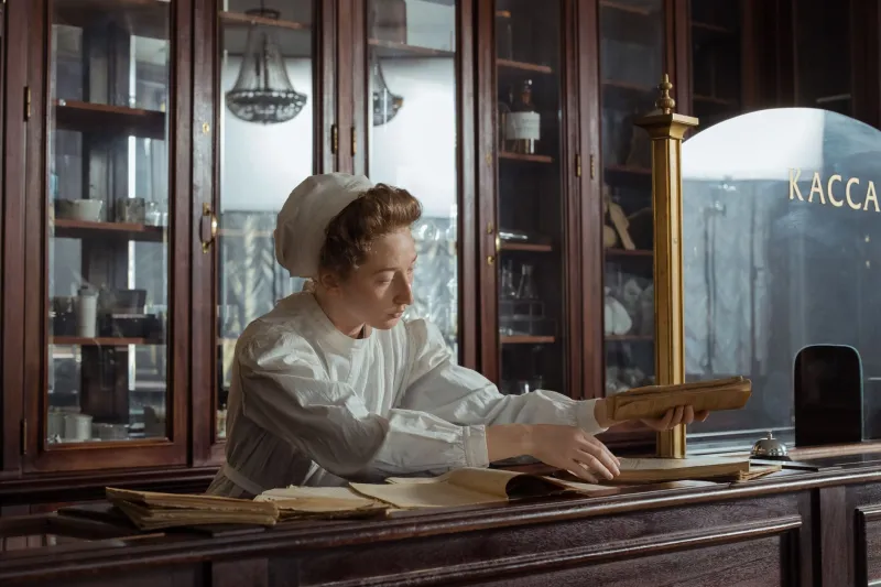
Imagine breaking your wrist in 1920. You'd show up at a doctor's office — maybe a general practitioner who also handled births, infections, and everything in between — and he'd press his fingers along your arm, feel for where things didn't line up, and make a judgment call. No images. No confirmation. Just experience, instinct, and a plaster cast applied over whatever position he thought was close enough.
For millions of Americans across the late 19th and early 20th centuries, that was the full extent of fracture care. Skilled physicians could do remarkable things with their hands alone. But remarkable wasn't the same as reliable. Bones set slightly off-angle would heal that way permanently. Fragments that weren't properly identified might shift inside the cast, causing complications that didn't show up until weeks later — when it was too late to easily correct them.
The result? A lot of crooked fingers. A lot of limps. A lot of wrists that ached every time the weather changed.
The Moment Everything Changed
Wilhelm Röntgen accidentally discovered X-rays in 1895, and within a year, American physicians were experimenting with the technology. It was a genuine revolution — suddenly, for the first time in history, a doctor could see inside a living body without cutting it open.
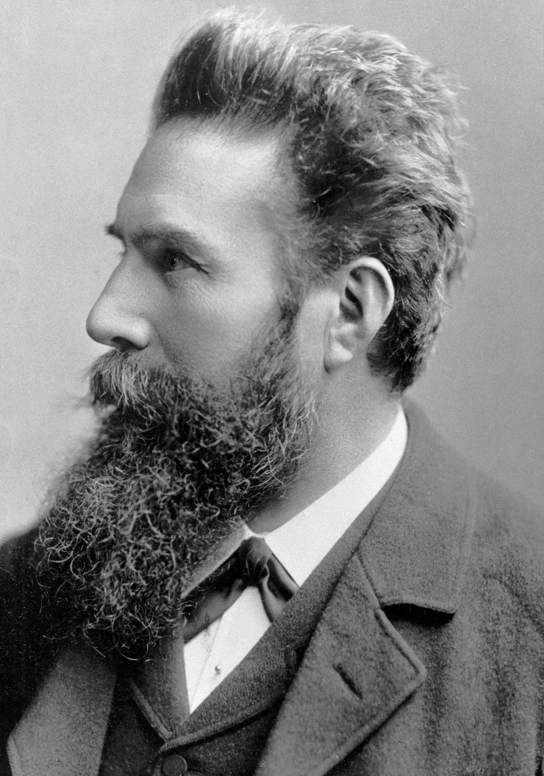 Photo: Wilhelm Röntgen, via cdn.britannica.com
But adoption was slow and uneven. Early X-ray machines were expensive, cumbersome, and concentrated in large urban hospitals. A farmer in rural Ohio with a broken leg in 1910 wasn't getting an X-ray. Neither was a factory worker in a small Pennsylvania town in 1930. The technology existed, but access was a different matter entirely.
Through the mid-20th century, X-ray use gradually spread. By the 1950s and 60s, most hospitals had the equipment. By the 1970s, it was becoming genuinely routine. And with better imaging came better outcomes — doctors could now see exactly where a bone had fractured, how many pieces were involved, and whether the break was clean or complex.
That single shift — from feeling to seeing — changed the entire logic of fracture care.
From Plaster and Hope to Surgical Hardware
Once physicians could properly visualize a fracture, treatment options expanded dramatically. A clean break in a forearm? A properly positioned cast, confirmed by imaging, with a follow-up X-ray to ensure nothing had shifted. A more complex fracture with multiple fragments? Surgical intervention, with titanium plates and screws anchoring the bone in exactly the right position while it healed.
The introduction of orthopedic hardware — metal rods, pins, plates, and screws — transformed what was possible. Fractures that once meant months of immobilization, permanent deformity, or even amputation could now be surgically repaired and stabilized with precision. The hardware did the work the cast used to do, but far more reliably.
By the 1980s and 90s, the field had advanced to the point where complex fractures were being repaired in dedicated orthopedic surgery centers rather than general hospital wards. Specialists who spent their entire careers focused on bones and joints replaced the generalists who had once handled everything.
CT Scans and the Second Revolution
If X-rays were the first revolution, CT scanning — computed tomography — was the second. Introduced in clinical settings in the 1970s and widely available by the 1980s and 90s, CT scans gave physicians a three-dimensional view of a fracture that flat X-rays simply couldn't provide.
For complex injuries — fractures near joints, spinal injuries, pelvic breaks — this mattered enormously. A CT scan could reveal exactly how fragments were positioned relative to each other, allowing surgeons to plan their approach before making a single incision. What used to require exploratory surgery could now be mapped out on a screen the night before the procedure.
MRI technology added another layer, allowing soft tissue — tendons, ligaments, cartilage — to be evaluated alongside the bone itself. A fractured ankle, for instance, might involve ligament damage that an X-ray would completely miss. MRI caught what everything else couldn't.
A Broken Wrist in 2025
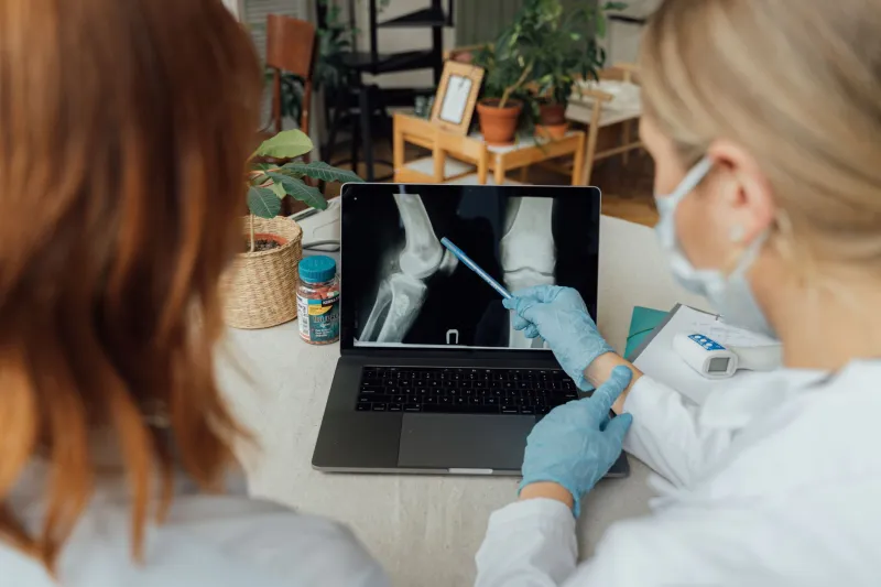
Here's what that same wrist fracture looks like today. You walk into an urgent care center or emergency room. Within minutes, a digital X-ray — far faster and lower-radiation than its predecessors — confirms the break and shows its exact nature. If it's a clean, stable fracture, you're fitted with a modern splint or lightweight cast and sent home with a follow-up appointment.
If it's more complex, an orthopedic surgeon reviews the imaging, often the same day. Surgery, if needed, is frequently performed as an outpatient procedure — you arrive in the morning, a surgeon places a titanium plate and a handful of screws under fluoroscopic guidance (real-time X-ray imaging), and you're home by afternoon. Physical therapy begins within weeks. Full function, in many cases, is restored within two to three months.
The entire process that once took six months of guesswork and left patients with permanent limitations now routinely ends with complete recovery.
What We Don't Fully Appreciate
There's something easy to overlook in all of this. Fractures are so common — so casually treatable today — that most people don't register them as serious medical events. A broken collarbone keeps a kid out of Little League for a few weeks. A fractured wrist means a few months of inconvenience. We've normalized outcomes that would have seemed extraordinary to any physician practicing before 1950.
The real story of X-rays and orthopedic imaging isn't just about technology. It's about the quiet elimination of a category of permanent suffering that used to be considered an unavoidable part of life. Crooked bones, chronic joint pain, limbs that never fully worked again — these weren't rare tragedies. They were common experiences, accepted as the natural consequence of injury.
We stopped accepting them so gradually that most of us never noticed the change.